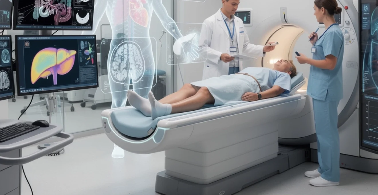
The revolution in medical imaging has fundamentally transformed how healthcare professionals approach disease detection, moving from reactive treatment to proactive prevention. Modern imaging technologies now possess the remarkable capability to identify pathological changes at the cellular and molecular level, often years before patients experience any noticeable symptoms. This paradigm shift represents one of the most significant advances in preventive medicine, offering unprecedented opportunities for early intervention and improved patient outcomes.
Advanced imaging modalities have evolved beyond simple structural visualisation to become sophisticated diagnostic tools that can detect metabolic changes, tissue composition alterations, and even genetic expressions. The integration of artificial intelligence and machine learning algorithms has further enhanced the sensitivity and specificity of these diagnostic techniques, enabling radiologists to identify subtle patterns that might otherwise remain undetected until disease progression reaches advanced stages.
Computed tomography screening protocols for asymptomatic disease detection
Computed tomography has emerged as a cornerstone of asymptomatic disease screening, offering exceptional spatial resolution and rapid acquisition times that make large-scale population screening programmes both feasible and cost-effective. The development of sophisticated CT protocols has enabled healthcare systems to implement comprehensive screening strategies that can detect various malignancies and cardiovascular conditions before clinical manifestation occurs.
The standardisation of CT screening protocols has been crucial in ensuring consistent results across different healthcare facilities and geographical regions. These protocols incorporate specific technical parameters including radiation dose optimisation, contrast timing protocols, and image reconstruction algorithms that maximise diagnostic accuracy whilst minimising patient exposure to ionising radiation.
Low-dose CT lung cancer screening in High-Risk populations
Low-dose computed tomography lung cancer screening has revolutionised early detection strategies for individuals at high risk of developing pulmonary malignancies. This screening modality utilises radiation doses approximately 90% lower than conventional chest CT examinations whilst maintaining diagnostic image quality sufficient for detecting lung nodules as small as 4-6 millimetres in diameter. The technique has demonstrated remarkable efficacy in reducing lung cancer mortality rates by up to 20% in high-risk populations, particularly among heavy smokers and individuals with significant occupational exposure to carcinogens.
The implementation of low-dose CT screening programmes requires careful patient selection based on established risk criteria, including age, smoking history, and family history of lung cancer. Advanced computer-aided detection systems now assist radiologists in identifying suspicious pulmonary nodules, whilst sophisticated algorithms help differentiate between benign and potentially malignant lesions based on morphological characteristics, growth patterns, and density measurements.
Coronary artery calcium scoring using electron beam CT
Coronary artery calcium scoring represents a non-invasive method for assessing cardiovascular risk in asymptomatic individuals through the quantification of calcified atherosclerotic plaques within the coronary arteries. This technique utilises specialised electron beam computed tomography or multi-detector CT scanners to generate detailed images of the heart during a brief breath-hold, typically lasting 10-15 seconds. The resulting calcium score provides valuable prognostic information that helps clinicians stratify patients into different cardiovascular risk categories.
The clinical significance of coronary calcium scoring extends beyond simple risk stratification, as it enables personalised therapeutic interventions based on individual patient risk profiles. Studies have demonstrated that patients with elevated calcium scores benefit significantly from aggressive lipid-lowering therapies and lifestyle modifications, even when traditional cardiovascular risk factors appear within normal ranges.
Hepatocellular carcinoma detection through Triple-Phase CT imaging
Triple-phase computed tomography imaging has become the gold standard for detecting hepatocellular carcinoma in high-risk patients, particularly those with chronic liver disease or cirrhosis. This sophisticated imaging protocol involves the administration of intravenous contrast material followed by image acquisition during three distinct phases: arterial, portal venous, and delayed phases. Each phase provides unique information about vascular perfusion patterns that help differentiate malignant lesions from benign liver abnormalities.
The characteristic enhancement patterns observed during triple-phase CT examinations enable radiologists to identify hepatocellular carcinomas as small as 1-2 centimetres in diameter. Early detection through this technique has been associated with significantly improved treatment outcomes, as patients diagnosed with small, localized tumours are often candidates for curative surgical resection
Additionally, multiphasic protocols allow the detection of subclinical vascular invasion and satellite nodules, factors that critically influence transplant eligibility and locoregional treatment planning. For asymptomatic patients enrolled in chronic liver disease surveillance programmes, triple-phase CT provides a powerful complement to ultrasound and laboratory markers, supporting truly pre-symptomatic diagnosis and timely intervention.
CT colonography virtual endoscopy techniques for colorectal cancer
CT colonography, often referred to as virtual colonoscopy, has emerged as an effective alternative to conventional optical colonoscopy for colorectal cancer screening in asymptomatic individuals. Using low-dose CT protocols and advanced 2D and 3D reconstruction algorithms, this technique generates high-resolution images of the colonic lumen, allowing radiologists to visualise polyps and early neoplastic lesions without the need for sedation or invasive instrumentation. For many patients who are reluctant to undergo traditional colonoscopy, CT colonography offers a more acceptable option that can significantly increase screening uptake.
From a technical perspective, virtual endoscopy relies on meticulous bowel preparation, colonic insufflation, and dual-position scanning to minimise blind spots and enhance polyp detection. Sophisticated software enables “fly-through” navigation of the colon, similar to driving a miniature camera through the bowel, while also allowing evaluation of extracolonic organs for incidental but clinically relevant findings. Early-stage colorectal cancers and advanced adenomas identified on CT colonography can then be targeted for therapeutic colonoscopy, enabling removal before symptoms such as bleeding, pain, or altered bowel habits develop.
Magnetic resonance imaging biomarkers in pre-clinical disease states
Magnetic resonance imaging has evolved into a powerful platform for detecting subtle tissue changes that precede overt structural damage, making it indispensable for identifying pre-clinical disease states. Unlike CT, MRI does not rely on ionising radiation, instead using strong magnetic fields and radiofrequency pulses to interrogate tissue composition, perfusion, and microstructure. Advanced MRI biomarkers now allow clinicians to detect disease processes such as inflammation, demyelination, fibrosis, and protein deposition years before patients present with clinical symptoms.
These quantitative MRI techniques generate reproducible measurements that can be tracked over time, providing objective markers of disease progression and therapeutic response. In many ways, MRI biomarkers function like a “laboratory test for tissues”, revealing early pathology invisible to conventional imaging. As we increasingly integrate MRI into preventive health strategies, asymptomatic individuals at genetic or lifestyle-based risk can benefit from tailored surveillance protocols that focus on the earliest detectable signs of disease.
Diffusion-weighted imaging for early stroke detection
Diffusion-weighted imaging (DWI) has become the cornerstone of ultra-early stroke detection, capable of identifying ischaemic changes within minutes of arterial occlusion. By measuring the random Brownian motion of water molecules within brain tissue, DWI reveals areas where diffusion is restricted due to cytotoxic oedema, often long before structural changes appear on conventional T1- or T2-weighted sequences. For patients who wake with subtle symptoms or present with ambiguous neurological signs, DWI can provide a definitive answer when time-critical treatment decisions must be made.
In the context of pre-symptomatic or minimally symptomatic disease, DWI is particularly valuable for detecting “silent” infarcts and transient ischaemic attacks that might otherwise go unrecognised. These clinically covert events are now understood to significantly increase the risk of subsequent major stroke and cognitive decline. By revealing these early lesions, diffusion MRI enables clinicians to intensify risk-factor management, optimise antithrombotic therapy, and implement lifestyle interventions before catastrophic neurological deficits develop.
Cardiac MRI T1 mapping for myocardial fibrosis assessment
Cardiac MRI T1 mapping has transformed our ability to detect diffuse myocardial fibrosis, a key driver of heart failure and arrhythmias that often develops long before patients notice shortness of breath or reduced exercise tolerance. Traditional imaging methods can miss early fibrotic change because the heart may still appear structurally normal, but T1 mapping quantifies the intrinsic relaxation properties of myocardial tissue, providing a sensitive biomarker of extracellular matrix expansion. When combined with contrast-enhanced techniques to calculate extracellular volume fraction, clinicians gain a detailed picture of both focal and diffuse fibrotic burden.
This quantitative approach allows subtle myocardial involvement to be detected in conditions such as hypertensive heart disease, diabetic cardiomyopathy, and inherited cardiomyopathies, even when ejection fraction remains preserved. For asymptomatic individuals with strong family histories or genetic variants associated with cardiomyopathy, T1 mapping facilitates earlier diagnosis and closer monitoring. In practical terms, this means therapy can be initiated while the myocardium is still relatively healthy, improving the chances of halting or reversing disease progression.
Brain amyloid imaging using FLAIR sequences in alzheimer’s disease
Fast fluid-attenuated inversion recovery (FLAIR) sequences play a pivotal role in the early imaging assessment of neurodegenerative disorders, including the pre-clinical phases of Alzheimer’s disease. While definitive amyloid imaging relies on PET radiotracers, FLAIR MRI is particularly sensitive to white matter hyperintensities, hippocampal signal changes, and subtle cortical alterations that often accompany vascular contributions to cognitive impairment. These changes may precede noticeable memory loss by several years, flagging individuals who would benefit from more comprehensive cognitive and biomarker evaluation.
In a preventative context, combining FLAIR imaging with volumetric MRI and advanced post-processing allows detection of early hippocampal atrophy and microstructural changes in key memory networks. You can think of this as seeing the “footprints” of amyloid and tau pathology in the brain’s architecture before clinical dementia emerges. Identifying such changes in asymptomatic but high-risk individuals—for example, those with strong family histories or genetic susceptibility—opens the door to intensified cardiovascular risk management, cognitive training, and potential enrolment in disease-modifying therapy trials.
Prostate-specific membrane antigen MRI for cancer screening
Prostate-specific membrane antigen (PSMA)-targeted imaging has gained prominence in recent years, and hybrid approaches that integrate PSMA-targeted contrast mechanisms with multiparametric MRI are reshaping early prostate cancer detection. Multiparametric MRI, which combines T2-weighted imaging, diffusion-weighted imaging, and dynamic contrast-enhanced sequences, already offers high sensitivity for clinically significant prostate tumours. When augmented with PSMA-based targeting strategies, radiologists can more confidently distinguish indolent lesions from those likely to progress, even when prostate-specific antigen (PSA) levels are only mildly elevated.
For asymptomatic men with borderline or slowly rising PSA, PSMA-enhanced MRI can reduce unnecessary biopsies by improving lesion characterisation and guiding targeted sampling. This tailored approach supports risk-adapted screening, where you are more likely to receive further investigation only when imaging biomarkers suggest a meaningful risk of aggressive disease. In the longer term, integrating PSMA MRI into prostate cancer screening protocols may help strike a better balance between early detection and overdiagnosis, ensuring that clinically important cancers are identified while avoiding overtreatment of low-risk lesions.
Ultrasound elastography applications in subclinical pathology
Ultrasound elastography has extended the capabilities of conventional sonography by providing real-time information about tissue stiffness, a property that often changes early in disease processes. By measuring how tissues deform in response to mechanical stress or acoustic forces, elastography generates colour-coded maps that highlight areas of increased or decreased elasticity. Since many conditions—such as fibrosis, malignancy, and chronic inflammation—alter tissue stiffness before size or morphology change, elastography is ideally suited for detecting subclinical pathology in asymptomatic patients.
From a practical standpoint, ultrasound elastography is relatively inexpensive, widely available, and free from ionising radiation, making it a valuable tool for longitudinal surveillance programmes. For patients, the experience is similar to a standard ultrasound examination, yet the information obtained is far more nuanced. In many ways, elastography can be compared to “palpation with superhuman sensitivity”, enabling clinicians to feel what their hands cannot in deep or small organs.
Shear wave elastography for liver fibrosis quantification
Shear wave elastography (SWE) has become an essential technique for non-invasive assessment of liver fibrosis, reducing the need for diagnostic biopsy in many patients with chronic liver disease. By generating and tracking the propagation speed of shear waves through hepatic tissue, SWE provides quantitative measurements of stiffness that correlate closely with fibrosis stage. Importantly, these measurements can detect significant fibrosis long before patients develop clinical signs such as jaundice, ascites, or variceal bleeding.
For individuals with risk factors like viral hepatitis, alcohol misuse, non-alcoholic fatty liver disease, or metabolic syndrome, shear wave elastography enables routine monitoring of liver health as part of a comprehensive preventative strategy. Serial measurements help clinicians evaluate response to lifestyle modifications and pharmacological therapy, highlighting improvements or progression at a stage when interventions are most effective. By transforming liver fibrosis into a trackable metric, SWE helps shift the focus from late-stage cirrhosis management to early disease modification.
Acoustic radiation force impulse imaging in thyroid nodule assessment
Acoustic radiation force impulse (ARFI) imaging is a specialised elastography technique that has significantly improved the evaluation of thyroid nodules detected incidentally on ultrasound or other imaging studies. ARFI uses short, high-intensity acoustic pulses to generate localised tissue displacement, from which stiffness values are calculated. Since malignant thyroid nodules often exhibit higher stiffness than benign ones, ARFI provides an additional diagnostic parameter beyond conventional ultrasonographic features such as echogenicity, margins, and calcifications.
In asymptomatic patients with incidentally discovered nodules, ARFI can help refine risk stratification and reduce the number of unnecessary fine-needle aspirations. When combined with established risk-scoring systems, elastography parameters support more confident decisions about which nodules warrant biopsy and which can be safely monitored. For patients, this means fewer invasive procedures and less anxiety, while still maintaining a high level of vigilance for clinically significant thyroid cancers.
Strain elastography techniques for breast cancer detection
Strain elastography has become a valuable adjunct to conventional breast ultrasound for distinguishing benign from malignant lesions in women who may have no palpable abnormalities. By evaluating tissue deformation in response to gentle compression or physiological motion, strain imaging produces qualitative and semi-quantitative assessments of lesion stiffness. Malignant breast tumours tend to be firmer and less compressible than surrounding tissue, and these differences are clearly visualised on elastographic maps.
For women undergoing screening—particularly those with dense breast tissue where mammography may be less sensitive—adding elastography to ultrasound can improve diagnostic accuracy and reduce false-positive findings. This is especially relevant for asymptomatic individuals referred after suspicious screening results, where the key question is often whether a lesion requires biopsy. By providing an additional layer of functional information, strain elastography supports more precise decision-making, helping to detect aggressive cancers early while minimising over-investigation of benign findings.
Positron emission tomography radiopharmaceuticals for molecular disease detection
Positron emission tomography (PET) offers a unique window into the molecular and metabolic processes that underlie disease, often revealing abnormalities long before structural imaging changes become apparent. Using radiopharmaceuticals that target specific receptors, enzymes, or metabolic pathways, PET can detect early tumour metabolism, neurodegeneration, and inflammatory activity in otherwise normal-appearing tissues. In the context of pre-symptomatic disease detection, this capability is particularly powerful, as it allows clinicians to identify and quantify disease activity at its earliest biochemical stages.
The most widely known tracer, 18F-FDG, highlights areas of increased glucose metabolism and has been central to oncology imaging for decades. However, a new generation of PET radiopharmaceuticals is rapidly expanding the range of detectable conditions, from amyloid and tau deposition in Alzheimer’s disease to PSMA expression in prostate cancer and somatostatin receptors in neuroendocrine tumours. As these targeted agents become more widely available, asymptomatic individuals at high risk—for example, due to genetic mutations or strong family histories—may benefit from selectively deployed PET imaging to guide surveillance and early intervention.
Artificial intelligence integration in medical imaging workflows
Artificial intelligence has become deeply integrated into modern medical imaging workflows, acting as a powerful assistant that augments, rather than replaces, the expertise of radiologists. By rapidly analysing large volumes of imaging data, AI algorithms can detect subtle patterns, quantify complex features, and flag abnormalities that might be challenging to discern with the naked eye. This is especially important in the context of early disease detection, where the imaging signs can be extremely subtle and easily overlooked amidst heavy workloads.
AI tools are now being deployed at multiple stages of the imaging pipeline, from optimising scan acquisition parameters to automating lesion segmentation and generating structured reports. For example, deep learning models can automatically calculate coronary artery calcium scores, classify lung nodules on low-dose CT, or measure brain volumes on MRI, providing consistent and reproducible metrics across time and institutions. For patients, this translates into more reliable screening results, shorter reporting times, and a reduced likelihood that small but significant findings are missed.
Another key benefit of AI integration is intelligent triage. Algorithms can prioritise imaging studies with suspected critical findings—such as intracranial haemorrhage, large vessel occlusion, or pneumothorax—so that they are reviewed first by radiologists. In emergency and stroke care, where every minute counts, this prioritisation can directly impact outcomes. Looking ahead, as we ask how medical imaging helps diagnose diseases before symptoms appear, AI is likely to be an essential component, enabling us to detect earlier, act faster, and personalise follow-up with unprecedented precision.
Radiogenomics and precision medicine through advanced imaging analytics
Radiogenomics is an emerging field that links imaging features with genomic and molecular characteristics of disease, providing a bridge between what we see on scans and what is happening at the cellular level. By applying advanced image analytics and machine learning to large datasets, researchers have identified imaging phenotypes that correlate with specific gene expression patterns, mutation profiles, and tumour microenvironment characteristics. In essence, radiogenomics aims to turn routine imaging into a non-invasive “virtual biopsy” that captures the molecular landscape of disease throughout the entire organ.
In oncology, this approach is particularly promising for tumours that are heterogeneous or difficult to access surgically. Imaging-based signatures can help predict tumour aggressiveness, likelihood of treatment response, and risk of metastasis long before clinical progression becomes apparent. For asymptomatic patients identified through screening—such as those with early lung nodules, small renal masses, or low-volume prostate cancer—radiogenomic markers may soon guide decisions about surveillance versus immediate intervention, supporting truly personalised care.
Beyond cancer, radiogenomics is being explored in cardiovascular, neurological, and inflammatory diseases, where imaging patterns are linked to pathways involved in fibrosis, neurodegeneration, and immune activation. As these insights mature, you can expect medical imaging to play an even larger role in precision medicine, not only diagnosing diseases before symptoms appear but also predicting how they will behave and which therapies will be most effective. Ultimately, the integration of radiogenomics, AI, and advanced imaging biomarkers heralds a future where preventive healthcare is tailored to each individual’s unique biological profile, long before illness has the chance to take hold.